

Admittedly, I have a love-hate relationship with technology. I enjoy using my smart phone and all the apps, but I have been known to cause computers, printers, and a robot or two to go haywire (maybe it’s my magnetic personality). But when it comes to brain-related technology and its groundbreaking applications, it is all love!
Some technologies allow us to study the structures of the brain, while others allow us to examine the functions of brain activity. I could spin off for hours discussing this exciting, and ever-developing area of research and the potential for my work with clients, but I will model "self-inhibition" and stick to the basics of neuro technology.
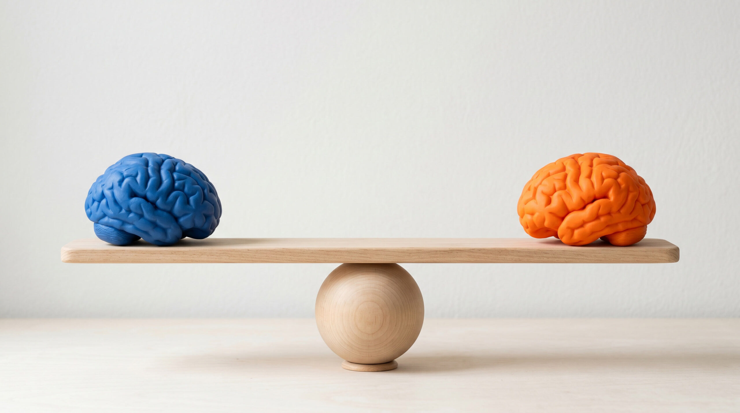
Neuro Technology - Looking Into the Brain
Most people by now have heard of Computerized Axial Tomography, also known as a CAT or CT scan, as well as Magnetic Resonance Imaging, which most people simply call an MRI. Without going into all the details of how these technologies work, suffice it to say that these are some of the best diagnostic tools the medical community currently has for finding tumors, deformities, and damages to the body and brain.
But when we want to learn more about how the brain works, the medical community has a hodgepodge of acronyms to toss in our medical charts. The four "biggies" of neuro technology include:
- Electroencephalography, also known as an EEG;
- Functional Magnetic Resonance Imaging, which we all call an fMRI
- Magnetoencephalography, which doctors call a MEG;
- Positron Emission Tomography, commonly termed a PET
What Brain Scans Tell Us
Again, without spinning out of control with my enthusiasm for "neuro technology", here's what the technologies basically allow. With the EEG and MEG, we can figure out how fast something happens in the brain by measuring electrical and magnetic activity. The activity is recorded on a computer using electrodes or magnetic detectors attached to the patient's scalp. Although the patient might look like a space alien during the session, he or she has no exposure to radiation.
A PET scan is a little more invasive. Doctors inject a radioactive substance into the patient. It goes through the brain and shows where levels of radiation accumulate. The patient wears a bunch of detectors that show areas of accumulation. The information creates a color image, with "hot spots" appearing red or orange and "quiet zones" blue or green. Generally, this procedure is not performed on children because of the radioactive risk.
My favorite–at the moment–is the fMRI, which is noninvasive, painless, and does not involve radiation. It allows practitioners to observe brain activity, based on blood flow. When any part of your brain gets more active, it needs more oxygen and nutrients. Hemoglobin, which has iron and is magnetic, carries oxygen to your brain. Using a big magnet, doctors can compare areas of oxygenated hemoglobin to deoxygenated hemoglobin entering the brain. A computer then takes the information and uses colors to identify the areas of the brain receiving the most blood. Pretty amazing, huh?
I wish that all of us could "have our heads examined". Just like a yearly physical, we could all get a yearly "mental" and learn more about the three pound wonder that is our brain.
Make it a successful day!